
Such as beta-blockers, sodium channel blockers, and calcium channel blockers. type 2 IVCD 5 intraventricular conduction delay IVR 5 idioventricular rhythm Junctional.

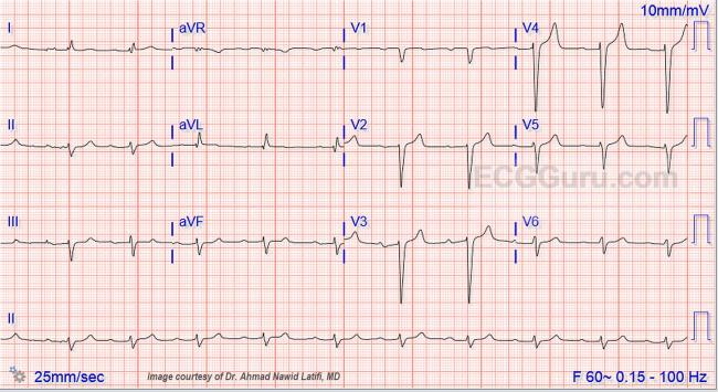
Sometimes, medications given to improve the heart’s rhythm can have the reverse effect and cause arrhythmias. The ECG: The rhythm is normal sinus at a rate of about 76 bpm with normal intervals. detection and classification of Sinus rhythm (Sinus), Atrial. The blocks can be total or partial (fascicular), accounting for a varied range of conduction. Concepts and types of IVCD Right bundle branch block. Medication side effects: Taking certain medications can impact a heart’s rate and rhythm. This is called intraventricular conduction delay (IVCD). Recognizing NSR Variations on NSR: Sinus arrhythmia Sinus bradycardia Sinus tachycardia. Conclusions: In a population study with long-term follow-up, NSIVCD and Minnesota definition of LBBB were independently associated with CV mortality. An EKG can reveal if the heart is beating out of rhythm or sequence. Background: Previous population studies have presented conflicting results regarding the prognostic impact of intraventricular conduction delays (IVCD). Heart rhythm abnormalities: A heart typically beats in a steady rhythm. Intraventricular conduction delay usually has no prognostic significance in patients without underlying heart disease but may progress to complete heart block or ventricular arrhythmia with worse prognosis in underlying. Natural History, Complications and Prognosis.

Ischemia, or lack of blood flow, may also cause an abnormal ECG. Bifascicular block (especially RBBB and LAF block) is the most common IVCD. This tissue will not conduct electricity as well, which can cause an abnormal ECG. Download scientific diagram Electrocargiogram showed Sinus rhythm (SR) with first degree AV block QRS with Intraventricular conduction defect (IVCD). Heart attack or ischemia: During a heart attack, blood flow in the heart is affected and heart tissue can begin to lose oxygen. In addition the ECG also shows an intraventricular conduction defect (IVCD) in the form of notched R wave in lead II and aVF and an rSR’ pattern in lead III. This occurs when the focus is in the low atrium (low atrial rhythm or coronary sinus rhythm). A superior P wave axis means that the atrial activation is proceeding from below upwards. If your electrolytes are imbalanced, you may have an abnormal ECG reading. ECG showing negative P waves in inferior leads II, III and aVF. These include:ĭefects or abnormalities in the heart’s shape and size: An abnormal ECG can signal that one or more aspects of the heart’s walls are larger than another meaning that the heart is working harder than normal to pump blood.Įlectrolyte imbalances: Electrolytes are electricity-conducting particles in the body that help keep the heart muscle beating in rhythm. Abnormal results can signify several issues.


 0 kommentar(er)
